Dissecting the Impact of Hepatocyte Inactivation Strategies on Drug Binding in Primary Hepatocyte Assays
Poster Authors:
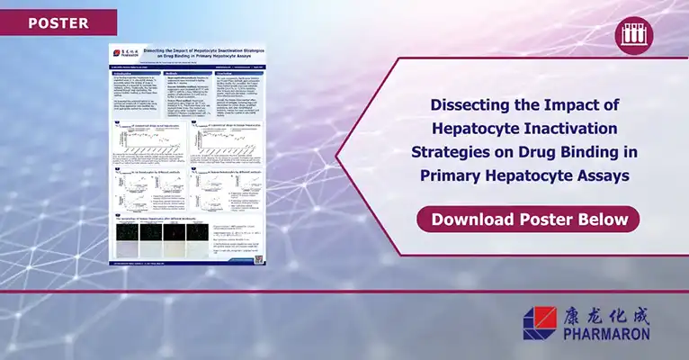
Xiaohui Chi, Rong Zhang, Jiafu Mu, Tong Li, Xinhao Xie, Yujie Duan, Chunyan Han, Man Xu
Pharmaron Beijing Co., Ltd. (China)
Hepatocyte assays and drug binding
Hepatocyte assays shape early ADME calls, and drug binding drives the unbound fraction that reaches targets. This overview explains why inactivation comes first, how common strategies work, and where each one fits. Then grab the poster to see the head-to-head data, images, and the exact steps used in the study.
What “drug binding in hepatocytes” really measures
When you measure binding in hepatocytes you want fu, the unbound fraction available to diffuse, transport, or clear. If metabolic enzymes keep working during the assay the result blends binding with turnover. That skews fu and clouds structure–activity trends. The fix is simple in principle. Stop metabolism cleanly before you measure.
Three ways teams inactivate hepatocytes
Heat inactivation.
Heating cells is fast and low cost. It denatures enzymes across pathways and needs no special reagents. The risk is overdoing it. Harsh heat can change protein structure that also drives nonspecific binding, which can shift fu for sticky compounds. Use this when you need a quick screen or when reagents are limited. Validate with a small panel first.
Enzyme inhibitor protocol.
A cocktail such as a heme-oxygenase mechanism-based inhibitor paired with a broad amidase or UGT blocker targets major metabolic routes at 37 °C. This keeps cell architecture closer to native. It adds setup time and complexity and you must show the inhibitors do not interfere with your analytes or LC-MS. It is a good fit when you want near-physiologic handling and you already track inhibitor blanks.
Freeze–thaw cycling.
Repeated freezing and thawing disrupts membranes and inactivates enzymes without adding proteins or small-molecule blockers. It is simple, repeatable, and helps when you want to avoid inhibitor carryover. Because membranes open up, confirm that the change does not shift nonspecific binding for highly lipophilic or basic drugs.
Practical controls that raise confidence
- Run a short metabolic stability check on one or two probe drugs to confirm inactivation
- Include recovery controls to spot adsorption to plastic or matrix
- Compare at least two media types since salts and protein supplements can move fu
- Use brightfield or basic fluorescence imaging to verify membrane disruption after treatment
- Match species to your IVIVE plan since fu in rat and human can differ for basic or high logP compounds
How to pick a default method
If you value speed and a short bill of materials start with freeze–thaw since it offers clean enzyme shutdown without extra reagents. If you need native-like handling and you already run inhibitor blanks an inhibitor cocktail works well. Heat is fine for scouting studies but confirm it does not inflate nonspecific binding for cationic or very lipophilic chemotypes.
What you will get from the poster
- Side-by-side methods with exact settings you can drop into an SOP
- Microscopy panels that show how inactivation changes cell morphology
- Comparative binding results across multiple drugs to benchmark your own lab
- Notes on media selection and species that speed method transfer
Download the poster to see the full workflow, imaging, and comparative results. It will help you standardize hepatocyte assays for drug binding and defend fu decisions in internal reviews.